TTA Surgery for Cruciate Ligament Rupture
What is TTA Surgery for Cruciate Ligament Rupture?
TTA is the abbreviation for tibial tuberosity advancement a common surgical procedure used to treat cranial (or anterior) cruciate ligament rupture in the knee joints of dogs.
Cranial cruciate ligament rupture is the most common cause of hind limb lameness in dogs. As a result, TTA and other operations that involve altering the shape of the tibia are common orthopaedic surgical procedures performed in dogs in specialist orthopaedic practices.
Why would an TTA Surgery for Cruciate Ligament Rupture be recommended?
Following rupture of the cranial cruciate ligament, the knee becomes unstable. When the dog takes weight on the limb this instability allows the shin bone to move forward. The stifle (knee) feels as though it is ‘giving-way’ and this can cause the dog to appear severely lame.
TTA surgery aims to make the tibial plateau perpendicular to the patellar tendon and, in doing so, prevent the shin bone moving forwards. The knee then feels stable for the dog when weight-bearing, despite the fact that the ligament has been ruptured and not directly repaired.
Candidates for TTA surgery are dogs with a ruptured cranial cruciate ligament that have persistent lameness and stifle joint instability. Young dogs and those with rupture of both of their ligaments (bilateral disease) are particularly good candidates.
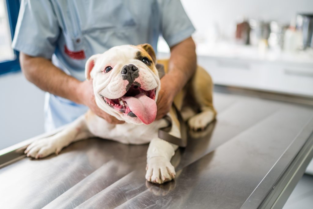
What does TTA Surgery for Cruciate Ligament Rupture involve?
Very specific X-rays need to be obtained of the stifle and tibia. The presence and severity of osteoarthritis can be assessed and the angle of the top of the shin bone measured. The position of the cut on the bone, the amount the bone needs to be rotated, and the size of plate necessary to stabilise the bone in its new position can then be evaluated.
Surgery may be performed on the same or a different day from the investigations. Prior to performing the TTA a small incision or cut is made into the knee joint to enable inspection of the structures within it.
Many dogs with ruptured cranial cruciate ligaments tear their cartilages (menisci). Damaged portions need to be removed. At the same time remnants of the ruptured ligament can be trimmed. A special implant is applied to the cut bone that has been designed especially for surgery.
X-rays are obtained at the end of the operation to assess the new angle of the top of the shin bone and check the position of the plate and screws.
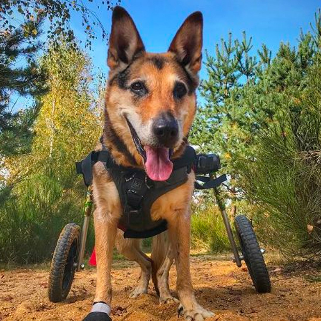
What is post surgical life like after TTA Surgery for Cruciate Ligament Rupture?
Aftercare following TTA surgery is very important, with rehabilitation taking many months. Exercise must be very restricted for the first few weeks until the soft tissues and cut bone heal, and at this stage is primarily for toileting purposes. It must be on a lead or harness to prevent strenuous activity. At other times confinement to a pen or a small room in the house is necessary with avoidance of jumping and climbing.
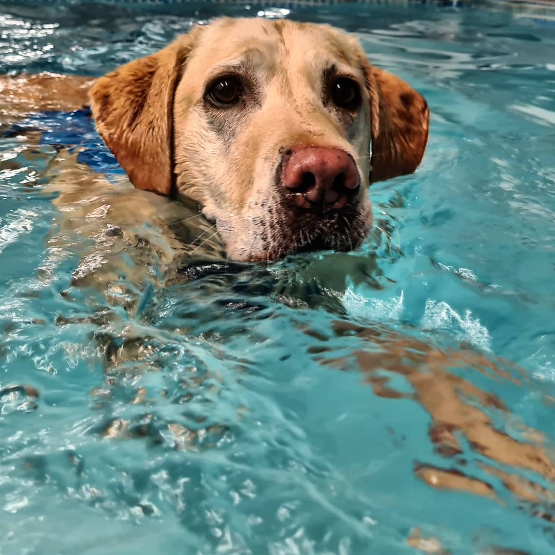
After a few weeks, exercise may be gradually increased in a controlled manner (still on a lead) as guided by a physiotherapist and Hydrotherapy may be recommended.
A check-up is necessary six to eight weeks after the operation. Limb and stifle function are checked at this time. X-rays are obtained to evaluate healing of the bone cut. Depending on progress advice is given regarding increasing exercise.
There are potential complications including infection, screw loosening and slow healing of the cut bone. A small percentage of dogs that didn’t have an injured cartilage at the time of TTA surgery tear it at a later date. A sudden increase in lameness usually develops and a second operation is necessary to remove the torn piece of cartilage.
However, although there is the potential for complications, in the majority of patients selected to undergo TTA surgery, knee pain is reduced and function of the limb is improved.
Post surgical rehabilitation
Rehabilitation is a process which aims to maximise patient mobility and wellbeing, returning them to their usual way of life following illness, injury or surgery. We restore pets to normal function (or as close as is possible), efficiently and safely using a wide variety of physiotherapeutic techniques.
Injury and even surgery can disrupt the body’s equilibrium in all sorts of direct and indirect ways. Even a pet’s own protective responses such as the inflammatory process can overwhelm and inhibit healing so one objective of rehabilitation is to reduce this level of inflammation. During rehabilitation, we also aim to boost the circulatory system, improve muscle function, increase range of motion within joints, and stimulate innate pain-relieving mechanisms.
With a committed and planned rehabilitation programme, pets can recover more quickly, realise better outcomes and avoid much pain and discomfort.
The best rehabilitation programmes consider the whole pet, not just the area of injury; we target and improve multiple systems throughout the body without forgetting the invaluable healing effects of boosting mental wellbeing too. From the wound healing properties of laser treatment, and the muscle strengthening of hydrotherapy, to the circulation boosting effects of massage, we will devise a rehabilitation programme to match a pet’s specific requirements.
Ready to get some help?
Our friendly and skilled physiotherapists are ready to help you and your dog with their rehabilitation.
More conditions
The content on this page is for advice and information only and does not represent veterinary guidance or direction. Please always consult a veterinary surgeon if you are worries about your dog.