Spondylosis deformans
What is Spondylosis deformans?
Spondylosis deformans is a condition that affects the vertebral bones of the spine and is best characterised by the presence of bony spurs or osteophytes along the edges of the bones of the spine.
A bony spur may develop in a single spot on the spine; more commonly, there will be multiple bone spurs in several different locations along the spine.
The most common places that spondylosis deformans lesions develop are along the thoracic vertebrae (chest), especially at the junction between the rib cage and the abdomen, in the lumbar spine (lower back) and in the lumbosacral spine (around the hips and back legs). In some cases the bony spurs may become large enough that they appear to form a complete bridge between adjacent vertebral bones.
Spondylosis deformans is a chronic condition that is associated with aging. Research indicates that it often develops as a secondary problem related to degenerative disease of the intervertebral discs.
What are the symptoms of Spondylosis deformans?
Most dogs with spondylosis deformans are free of any symptoms. Affected dogs may show signs of:
- Restrictive spinal movement
- May appear stiffer
- Pain in the back or hind legs
- Lameness in hind limbs
- Neurological delays
How is Spondylosis deformans diagnosed?
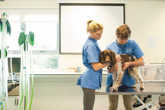
This condition is usually diagnosed from X-rays of the spine. In some cases, it may be an ‘incidental finding’ that is noticed when radiographs are taken for some other reason.
How is a Spondylosis deformans treated?
Treatment recommendations depend on the individual dog and whether or not it is showing any clinical signs.
Most dogs with spondylosis deformans appear to be pain-free and in these cases treatment is not necessary. If the pet appears to be painful, non-steroidal anti-inflammatory drugs (NSAIDs) or other analgesics may provide relief. Physiotherapy, weight loss, and controlled exercise programs may be helpful in some cases.
In rare cases, the osteophytes may be causing spinal cord compression, and in these cases, surgery to remove them may be indicated.
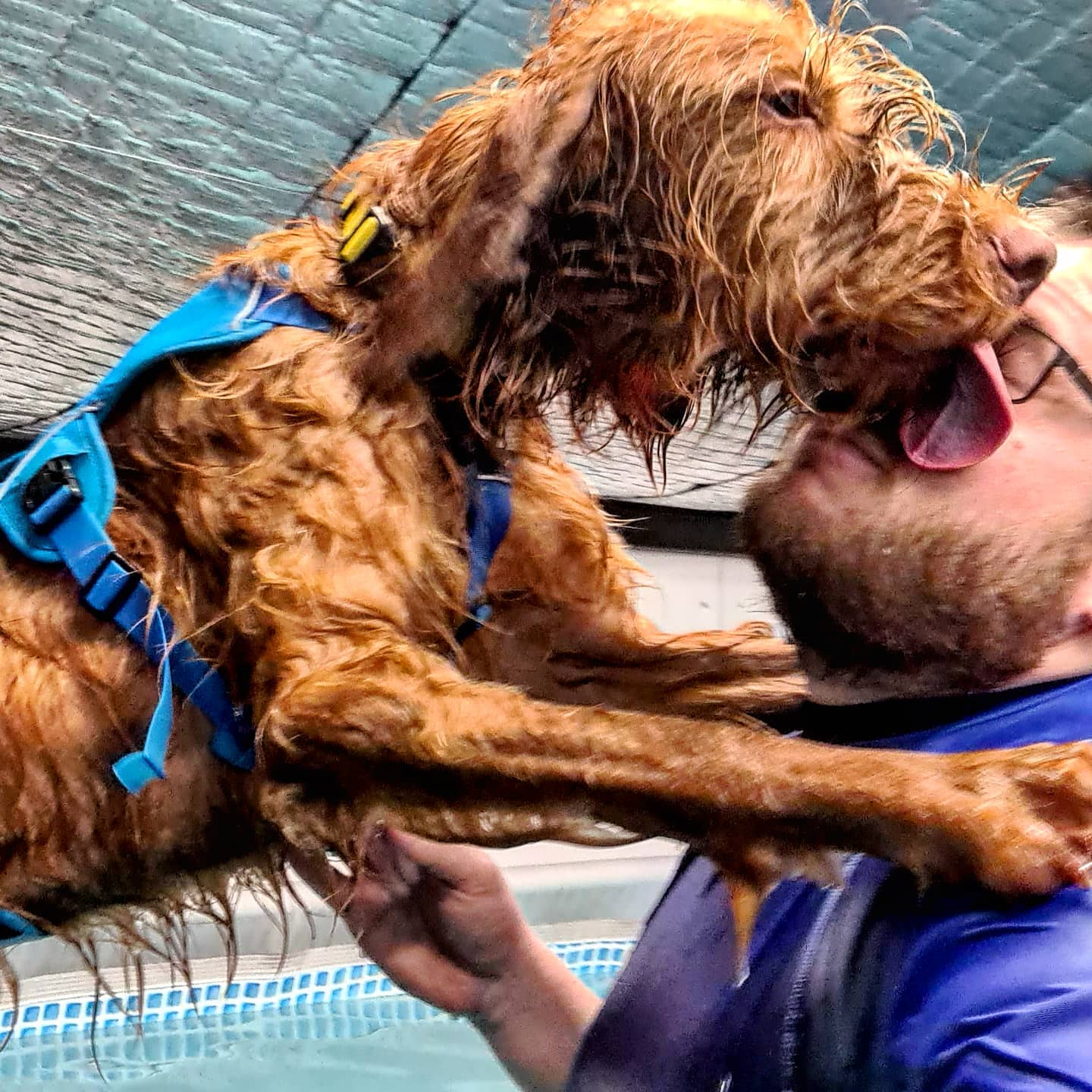
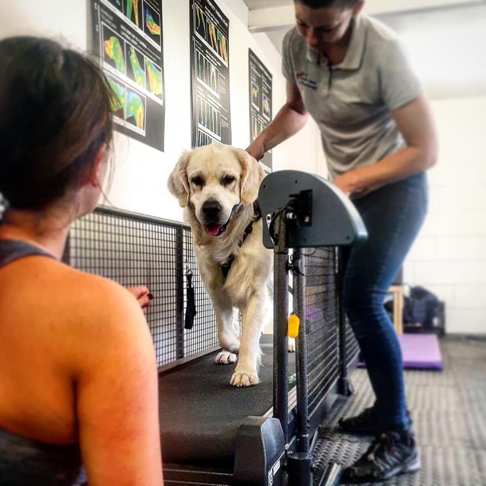
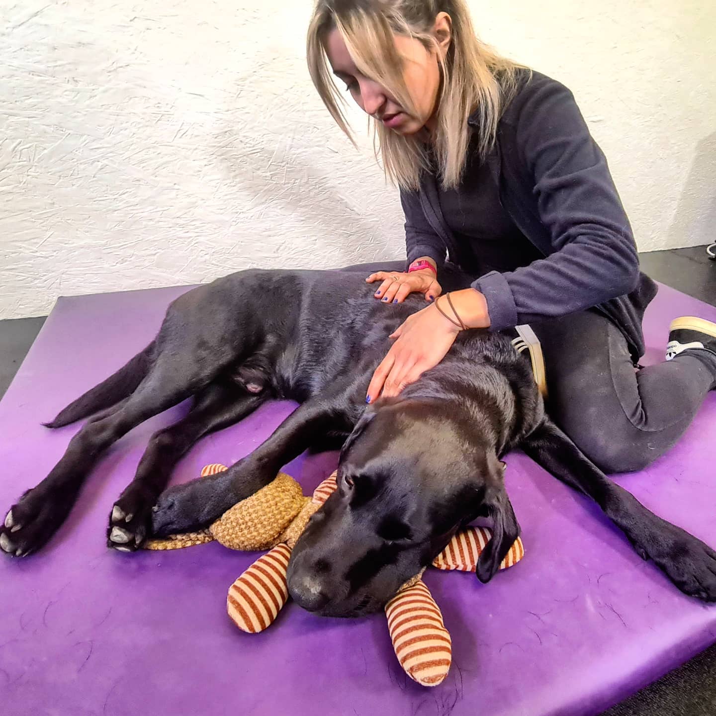
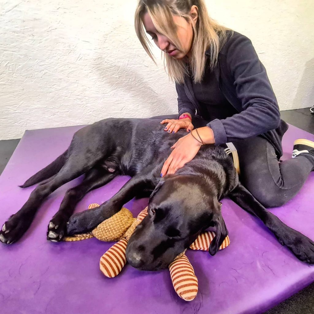
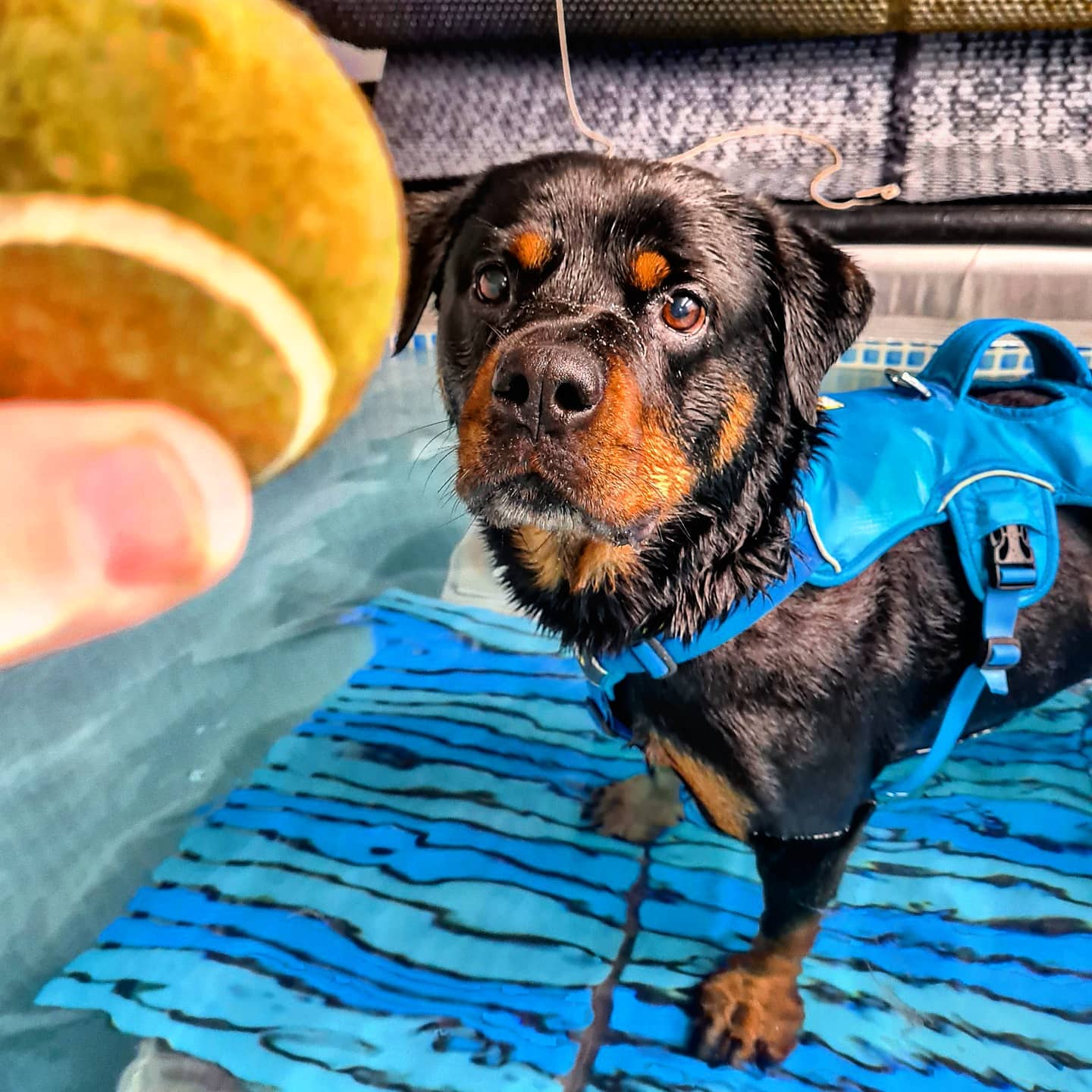
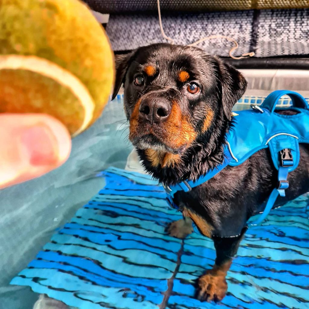
Orthopaedic rehabilitation
Rehabilitation is a process which aims to maximise patient mobility and wellbeing, returning them to their usual way of life following illness, injury or surgery. We restore pets to normal function (or as close as is possible), efficiently and safely using a wide variety of physiotherapeutic techniques.
Injury and even surgery can disrupt the body’s equilibrium in all sorts of direct and indirect ways. Even a pet’s own protective responses such as the inflammatory process can overwhelm and inhibit healing so one objective of rehabilitation is to reduce this level of inflammation. During rehabilitation, we also aim to boost the circulatory system, improve muscle function, increase range of motion within joints, and stimulate innate pain-relieving mechanisms.
With a committed and planned rehabilitation programme, pets can recover more quickly, realise better outcomes and avoid much pain and discomfort.
The best rehabilitation programmes consider the whole pet, not just the area of injury; we target and improve multiple systems throughout the body without forgetting the invaluable healing effects of boosting mental wellbeing too. From the wound healing properties of laser treatment, and the muscle strengthening of hydrotherapy, to the circulation boosting effects of massage, we will devise a rehabilitation programme to match a pet’s specific requirements.
Ready to get some help?
Our friendly and skilled physiotherapists are ready to help you and your dog with their rehabilitation.
More conditions
The content on this page is for advice and information only and does not represent veterinary guidance or direction. Please always consult a veterinary surgeon if you are worries about your dog.